[F1ANNEX VIU.K. METHODS OF ANALYSIS FOR THE DETERMINATION OF CONSTITUENTS OF ANIMAL ORIGIN FOR THE OFFICIAL CONTROL OF FEED
Textual Amendments
2. METHODS U.K.
2.1. Light microscopy U.K.
2.1.4. Microscopic examination U.K.
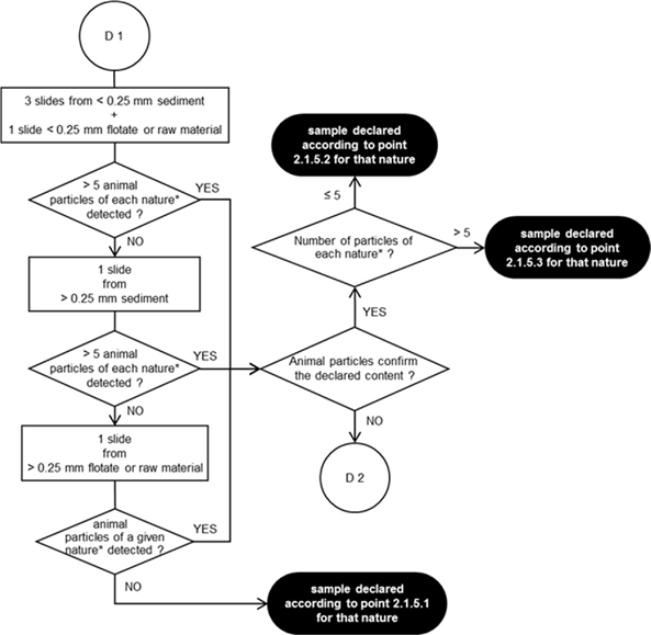
[F22.1.4.2. Observation flowchart for the detection of animal particles in compound feed and feed material U.K.
The prepared microscopic slides shall be observed in accordance with the observation flowcharts laid down in diagrams 1 and 2.
The microscopic observations shall be conducted using the compound microscope on the sediment and, depending on the operator’s choice, either on the flotate or on the raw material. The stereomicroscope may be used in addition to the compound microscope for the coarse fractions. Each slide shall be screened entirely at various magnifications. Precise explanations on how to use the observation flowcharts are detailed by a SOP established by the EURL-AP and published on its website.
The minimum numbers of slides to be observed at each step of the observation flowcharts shall be strictly respected, unless the entire fraction material does not permit to reach the stipulated slide number, for instance when no sediment is obtained. No more than 6 slides per determination shall be used for recording of the number of particles.
When additional slides are prepared on the flotate or the raw material using a more specific mounting medium with staining properties, as laid down in point 2.1.2.1.4, to further characterise structures (e.g. feathers, hairs, muscle or blood particles) which have been detected on slides prepared by other mounting media, as laid down in point 2.1.2.1.3, the number of particles shall be counted based on a number of slides per determination not exceeding 6, including the additional slides with a more specific mounting medium.
In order to facilitate the identification of the particles’ nature and origin, the operator may use support tools like decision support systems, image libraries and reference samples.]]

Textual Amendments